;void(0);)
By Jackie Trischman, Ph.D.
Long ago, if someone felt sick, doctors had to use their knowledge and experience to guess what was wrong. They had to rely on what they could see or feel to diagnose what was going on inside a patient’s body. But then a clever scientist named Wilhelm Conrad Roentgen had a breakthrough. He discovered a way to see inside the human body using X-rays, a special kind of light.
With his discovery, Roentgen created the first X-ray image of a human hand. It was like having a superpower!
X-rays are invisible to the human eye and pass right through skin and most organs. However, because bones are made of dense materials that contain calcium, they absorb X-rays and create shadows wherever they are blocked. Medical professionals use film or a special sensor (like in a digital camera) to detect and capture the X-rays as they pass through the body.
Using X-rays was a major improvement over guessing what was happening inside someone’s body. But X-rays also have limitations. As time passed, more brilliant minds joined the quest to improve medical imaging. They developed new types of scanners that let doctors easily see what was going on inside a patient, including:
- CT scanners — short for Computed Tomography, these provide 3-dimensional, detailed views of organs inside a patient’s body, made by moving an X-ray scanner around the body.
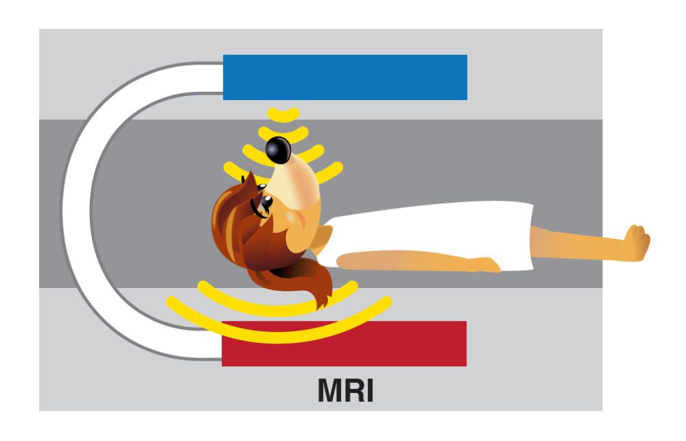
- MRIs — short for Magnetic Resonance Imaging, these machines use strong magnetic fields and radio waves to capture images of body parts that are not bones.
- Ultrasound — these use sound waves to create images that doctors and patients can look at to understand what’s going on inside organs and tissues in real time.
Each of these techniques has its own benefits, allowing doctors to see different parts of the body in amazing detail. They are used to detect and diagnose disease, check how well organs are working, and track how well a treatment is going. All this modern technology allows doctors to see much more than broken bones!
Scientists continue to dream big and push the boundaries of what is possible. Chemists work with computer scientists and physicists every day to invent new techniques and improve existing ones. As a result, they are making medical imaging even more powerful and precise.
Jackie Trischman, Ph.D. is Dean of the College of STEM at California State University, San Marcos.